Monday, May 8, 2017

Latest breast cancer discoveries
   The past twelve months were a fruitful time for breast cancer research. We hope the discoveries being made will provide hope and inspiration to all those living with breast cancer, and to the 17,586 women and 144 men expected to be diagnosed in 2017.

Featured here is BCNA’s top 10 list of breast cancer discoveries for 2016. We have chosen these discoveries for their contribution to breast cancer prevention, their breakthroughs in treatment outcomes and their contribution to the quality of life of Australians diagnosed with breast cancer.

Advances in prevention and early detection

1. Newly discovered rare genetic mutations linked to higher breast cancer risk

A global study has identified rare genetic changes that increase the risk of developing breast cancer. People who carry these rare mutations have been found to have a similar risk of developing breast cancer as those who carry the more common BRCA1 and BRCA2 gene mutations. Learning about rare genetic mutations is helpful for researchers so they can continue to investigate how best to reduce risk and treat people at high genetic risk.  

2. Osteoporosis drug may help prevent breast cancer in people at high risk

Cancer researchers at Walter and Eliza Hall Institute have found that denosumab (Xgeva), a drug most commonly used in osteoporosis treatment, is effective in preventing breast cancer in women who carry a BRCA1 gene mutation. This breakthrough may mean women with a BRCA1 gene mutation will have a less invasive option for reducing their risk of breast cancer in the future.

3. Longer-term use of aromatase inhibitors may help stop some breast cancers returning

Researchers in the US have found that taking the aromatase inhibitor letrozole for 10 years instead of five can further reduce the risk of breast cancer coming back for some women. Letrozole may also be effective in preventing new cancers from developing in the opposite breast.

4. Blood test may help predict when a cancer is returning

New research shows that a blood test might accurately predict the return of cancer in people who have been previously treated for early stage cancers. The blood test, also known as ‘liquid biopsy’, looks for cancer DNA in the bloodstream from cancer cells that have resisted treatment.


Advances in treatment

5. New targeted therapies for a common type of metastatic breast cancer

The PALOMA2 clinical trial found the new breast cancer drug palbociclib (Ibrance) to be an effective new first line treatment for hormone receptor positive, HER2-negative breast cancer when used in combination with the hormone therapy drug letrozole. The addition of palbociclib almost doubled progression free survival from 9 to 18 months.

A subsequent trial in later line therapies has found that  palbociclib used in combination with fulvestrant (Faslodex), can slow cancer growth in around two-thirds of women with hormone receptor positive, HER2-negative metastatic breast cancer.

6. New targeted treatments for triple negative breast cancer

Three recent clinical trials have shown promising results in the treatment of triple negative breast cancer by using the immune system to target the growth of cancer cells.

7. Tykerb and Herceptin in the treatment of HER2-positive early breast cancer

New research shows that giving a combination of the targeted drugs trastuzumab (Herceptin) and lapatinib (Tykerb) before surgery is effective in rapidly shrinking some HER2-positive breast cancers.


Improvements in quality of life and management of side effects

8. Scalp cooling systems can help reduce hair loss during chemotherapy

Research has shown that a scalp cooling system can reduce severe hair loss by 50 per cent in some women who are going through chemotherapy.

9. New online interventions can help women manage fear of recurrence

An Australian study testing the effectiveness of one-on-one therapy sessions using new psychological techniques has found a 22 per cent reduction in the fear of breast cancer returning.

10. Research shows personalised exercise programs can help with treatment side effects in early breast cancer

An Edith Cowen University study has shown that personalised exercise programs can help reduce treatment side effects in early breast cancer. The research demonstrates that moderate, supervised exercise can increase energy levels, help with nausea and muscle loss, and may help some people to recover faster.

The findings were presented in an ABC Catalyst episode and can be viewed here.
 Symptoms and Diagnosis of Medullary Carcinoma of the Breast

Signs and symptoms of Medullary Carcinoma of the Breast

   Like other types of breast cancer, medullary carcinoma may not cause any symptoms at first. Over time, a lump can form, and it may be soft and fleshy or somewhat firm to the touch. Most medullary carcinomas are small — less than 2 cm in size. Medullary carcinoma also may cause pain, swelling, redness, or tenderness in the breast.


Diagnosis

Tests that obtain images of the tissue inside the breast, such as mammography and ultrasound, sometimes can find a medullary carcinoma. On these tests, medullary carcinoma appears as a small, well-defined lump. However, some research suggests that mammography is not reliable at detecting medullary carcinomas. One study showed that these cancers were more likely to be found during self-examination or examination of the breasts by a doctor.

Diagnosing medullary carcinoma usually involves a combination of steps:

  • A physical examination of the breasts. Your doctor may be able to feel the lump in the breast.
  • A mammogram to locate the tumor and check for evidence of cancer in other areas of the breast.
  • Ultrasound to obtain additional images of the breast and check for other areas of cancer.
  • Biopsy to remove some or all of the lump to look at it under the microscope. A few cells from the lump can be extracted using a special needle, or the lump can be removed through a small incision. Biopsy is the key to accurate diagnosis, because imaging tests alone can’t tell the difference between medullary carcinoma and other types of breast cancer.

Also Read :What is Medullary Carcinoma of the Breast
When looked at under a microscope, medullary carcinoma has a number of important features that a pathologist looks for:

  • A clear, well-defined boundary between tumor tissue and normal breast tissue. Medullary carcinoma pushes against the surrounding healthy tissue, but doesn’t grow into it in the same way invasive ductal carcinoma usually does.
  • Large-sized cancer cells with a high-grade appearance, meaning that they look very different from normal, healthy breast cells. However, medullary carcinoma cells do not behave like high-grade cancer cells, which are aggressive and grow and spread quickly. Medullary cancer cells also tend to blend together in a distinctive “sheetlike” pattern; it’s hard to see each cell’s individual membrane (outer lining).
  • Immune system cells (white blood cells called lymphocytes and plasma cells) at the edges of the tumor. Immune system cells work to fight off diseases and any substances they see as a threat to the body. It’s believed that these cells help keep the medullary carcinoma in check, preventing it from growing and spreading quickly.

If the tumor has all of these features, it is considered to be a “true” medullary carcinoma. Sometimes the tumor has only some of these features but not others, or there may be some invasive ductal carcinoma cells mixed in. In these cases, your doctor may call the tumor “atypical medullary carcinoma.”

In addition, medullary carcinoma cells often express a protein called p53. The pathologist may test for p53 to help decide if the cancer is truly medullary.

There are some other key features of medullary carcinoma:

  • Hormone-receptor-negative: Medullary carcinoma usually tests negative for hormone receptors.
  • HER2-negative: Medullary carcinoma also usually tests negative for receptors for the protein HER2/neu.
Diagnosing medullary carcinoma can be challenging. It may be hard to tell the difference between medullary carcinoma cells and cells that make up a usual invasive ductal carcinoma. If you receive a diagnosis of medullary carcinoma, ask your doctor whether the pathologist who looked at the sample has experience diagnosing this type of cancer. You also may want to get a second opinion from another hospital pathology lab.
Medullary Carcinoma of the Breast
Medullary carcinoma of the breast is a rare subtype of invasive ductal carcinoma (cancer that begins in the milk duct and spreads beyond it), accounting for about 3-5% of all cases of breast cancer. It is called “medullary” carcinoma because the tumor is a soft, fleshy mass that resembles a part of the brain called the medulla.

Medullary Carcinoma of the Breast-whatisbreastcancer.info

 Medullary carcinoma can occur at any age, but it usually affects women in their late 40s and early 50s. Medullary carcinoma is more common in women who have a BRCA1 mutation. Studies have shown that medullary carcinoma is also more common in Japan than in the United States.

Medullary carcinoma cells are usually high-grade in their appearance and low-grade in their behavior. In other words, they look like aggressive, highly abnormal cancer cells, but they don’t act like them. Medullary carcinoma doesn’t grow quickly and usually doesn’t spread outside the breast to the lymph nodes. For this reason, it’s typically easier to treat than other types of breast cancer.

Read : Symptoms and Diagnosis of Medullary Carcinoma of the Breast

Tuesday, May 2, 2017

Symptoms of DCIS

   
DCIS generally has no signs or symptoms. A small number of people may have a lump in the breast or some discharge coming out of the nipple. According to the National Cancer Institute, about 80% of DCIS cases are found by mammography.
Read Also : What is DCIS Breast Cancer
Read Also : Diagnosis of DCIS
Tubular Carcinoma of the Breast
Tubular carcinoma of the breast is a subtype of invasive ductal carcinoma (cancer that begins inside the breast's milk duct and spreads beyond it into healthy tissue). Tubular carcinomas are usually small (about 1 cm or less) and made up of tube-shaped structures called "tubules." These tumors tend to be low-grade, meaning that their cells look somewhat similar to normal, healthy cells and tend to grow slowly.
Tubular Carcinoma of the Breast
At one time, tubular carcinomas accounted for about 1-4% of all breast cancers. Now that screening mammography is widely used, however, tubular carcinomas are being diagnosed more frequently — often before you or your doctor would be able to feel a lump. Exact numbers aren't available, but studies suggest that tubular carcinomas may account for anywhere from just under 8% to 27% of all breast cancers.

Studies also suggest that the average age of diagnosis for tubular carcinoma is the early 50s, although women can be diagnosed with it at any age. This type of cancer is rare in men.

Even though tubular carcinoma is an invasive breast cancer, it tends to be a less aggressive type that responds well to treatment. It isn't likely to spread outside the breast and is considered to have a very good prognosis.

Friday, April 28, 2017

IDC — Invasive Ductal Carcinoma
Invasive ductal carcinoma (IDC), sometimes called infiltrating ductal carcinoma, is the most common type of breast cancer. About 80% of all breast cancers are invasive ductal carcinomas.

Invasive means that the cancer has “invaded” or spread to the surrounding breast tissues. Ductal means that the cancer began in the milk ducts, which are the “pipes” that carry milk from the milk-producing lobules to the nipple. Carcinoma refers to any cancer that begins in the skin or other tissues that cover internal organs — such as breast tissue. All together, “invasive ductal carcinoma” refers to cancer that has broken through the wall of the milk duct and begun to invade the tissues of the breast. Over time, invasive ductal carcinoma can spread to the lymph nodes and possibly to other areas of the body.

Invasive Ductal Carcinoma.IDC Breast Cancer

Normal breast with invasive ductal carcinoma (IDC) in an enlarged cross-section of the duct
Breast profile:
A Ducts
B Lobules
C Dilated section of duct to hold milk
D Nipple
E Fat
F Pectoralis major muscle
G Chest wall/rib cage
Enlargement
A Normal duct cell
B Ductal cancer cells breaking through the basement membrane.
C Basement membrane


According to the American Cancer Society, more than 180,000 women in the United States find out they have invasive breast cancer each year. Most of them are diagnosed with invasive ductal carcinoma.

Although invasive ductal carcinoma can affect women at any age, it is more common as women grow older. According to the American Cancer Society, about two-thirds of women are 55 or older when they are diagnosed with an invasive breast cancer. Invasive ductal carcinoma also affects men.

Wednesday, April 26, 2017

DCIS — Ductal Carcinoma In Situ
Ductal carcinoma in situ (DCIS) is the most common type of non-invasive breast cancer. Ductal means that the cancer starts inside the milk ducts, carcinoma refers to any cancer that begins in the skin or other tissues (including breast tissue) that cover or line the internal organs, and in situ means "in its original place." DCIS is called "non-invasive" because it hasn’t spread beyond the milk duct into any normal surrounding breast tissue. DCIS isn’t life-threatening, but having DCIS can increase the risk of developing an invasive breast cancer later on.


When you have had DCIS, you are at higher risk for the cancer coming back or for developing a new breast cancer than a person who has never had breast cancer before. Most recurrences happen within the 5 to 10 years after initial diagnosis. The chances of a recurrence are under 30%.

Women who have breast-conserving surgery (lumpectomy) for DCIS without radiation therapy have about a 25% to 30% chance of having a recurrence at some point in the future. Including radiation therapy in the treatment plan after surgery drops the risk of recurrence to about 15%. Learn what additional steps you can take to lower your risk of a new breast cancer diagnosis or a recurrence in the Lower Your Risk section. If breast cancer does come back after earlier DCIS treatment, the recurrence is non-invasive (DCIS again) about half the time and invasive about half the time. (DCIS itself is NOT invasive.)

According to the American Cancer Society, about 60,000 cases of DCIS are diagnosed in the United States each year, accounting for about 1 out of every 5 new breast cancer cases.

There are two main reasons this number is so large and has been increasing over time:

People are living much longer lives. As we grow older, our risk of breast cancer increases.
More people are getting mammograms, and the quality of the mammograms has improved. With better screening, more cancers are being spotted early.
On the following pages you can learn about:

Monday, April 24, 2017

Types of Breast Cancer
Breast cancer can begin in different areas of the breast — the ducts, the lobules, or in some cases, the tissue in between. In this section, you can learn about the different types of breast cancer, including non-invasive, invasive, recurrent, and metastatic breast cancers, as well as the intrinsic or molecular subtypes of breast cancer. You can also read about breast cancer in men.


  • DCIS — Ductal Carcinoma In Situ
  • IDC — Invasive Ductal Carcinoma
  • IDC Type: Tubular Carcinoma of the Breast
  • IDC Type: Medullary Carcinoma of the Breast
  • IDC Type: Mucinous Carcinoma of the Breast
  • IDC Type: Papillary Carcinoma of the Breast
  • IDC Type: Cribriform Carcinoma of the Breast
  • ILC — Invasive Lobular Carcinoma
  • Inflammatory Breast Cancer
  • LCIS — Lobular Carcinoma In Situ
  • Male Breast Cancer
  • Molecular Subtypes of Breast Cancer
  • Paget's Disease of the Nipple
  • Phyllodes Tumors of the Breast
  • Recurrent & Metastatic Breast Cancer
Symptoms of Breast Cancer
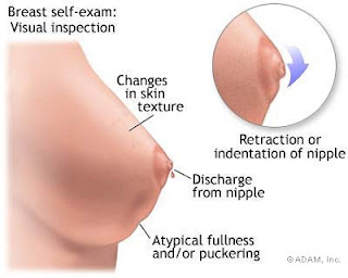
Initially, breast cancer may not cause any symptoms. A lump may be too small for you to feel or to cause any unusual changes you can notice on your own. Often, an abnormal area turns up on a screening mammogram (X-ray of the breast), which leads to further testing.

Symptoms of Breast Cancer
In some cases, however, the first sign of breast cancer is a new lump or mass in the breast that you or your doctor can feel. A lump that is painless, hard, and has uneven edges is more likely to be cancer. But sometimes cancers can be tender, soft, and rounded. So it's important to have anything unusual checked by your doctor.

According to the American Cancer Society, any of the following unusual changes in the breast can be a symptom of breast cancer:

swelling of all or part of the breast
skin irritation or dimpling
breast pain
nipple pain or the nipple turning inward
redness, scaliness, or thickening of the nipple or breast skin
a nipple discharge other than breast milk
a lump in the underarm area
These changes also can be signs of less serious conditions that are not cancerous, such as an infection or a cyst. It’s important to get any breast changes checked out promptly by a doctor.

In the following posts , you can learn about:

Understanding Breast Cancer

How breast cancer happens, how it progresses, the stages, and a look at risk factors.
Screening and Testing
The tests used for screening, diagnosis, and monitoring, including mammograms, ultrasound, MRI, CAT scans, PET scans, and more.

Types of Breast Cancer

The different types of breast cancer, including ductal carcinoma in situ (DCIS), invasive ductal carcinoma (IDC), invasive lobular carcinoma (ILC), inflammatory breast cancer, male breast cancer, recurrent breast cancer, metastatic breast cancer, and more.

Your Diagnosis

The characteristics of the cancer — featured on your pathology report — that might affect your treatment plan, including size, stage, lymph node status, hormone receptor status, and more.

Friday, April 21, 2017

Breast Cancer Risk and Risk Factors
You may be familiar with the statistic that says 1 in 8 women will develop invasive breast cancer. Many people misinterpret this to mean that, on any given day, they and the women they know have a 1-in-8 risk of developing the disease. That’s simply not true.
In reality, about 1 in 8 women in the United States — 12%, or about 12 out of every 100 — can expect to develop breast cancer over the course of an entire lifetime. In the U.S., an average lifetime is about 80 years. So, it’s more accurate to say that 1 in 8 women in the U.S. who reach the age of 80 can expect to develop breast cancer. In each decade of life, the risk of getting breast cancer is actually lower than 12% for most women.
People tend to have very different ways of viewing risk. For you, a 1-in-8 lifetime risk may seem like a high likelihood of getting breast cancer. Or you may turn this around and reason that there is a 7-in-8, or 87.5%, chance you will never get breast cancer, even if you live to age 80. How you view risk often depends on your individual situation — for example, whether you or many women you know have had breast cancer, or you have reason to believe you are at higher-than-normal risk for the disease — and your usual way of looking at the world.
Even though studies have found that women have a 12% lifetime risk of developing breast cancer, your individual risk may be higher or lower than that. Individual risk is affected by many different factors, such as family history, reproductive history, lifestyle, environment, and others.
What Is Breast Cancer?
Breast cancer is an uncontrolled growth of breast cells. To better understand breast cancer, it helps to understand how any cancer can develop.
Cancer occurs as a result of mutations, or abnormal changes, in the genes responsible for regulating the growth of cells and keeping them healthy. The genes are in each cell’s nucleus, which acts as the “control room” of each cell. Normally, the cells in our bodies replace themselves through an orderly process of cell growth: healthy new cells take over as old ones die out. But over time, mutations can “turn on” certain genes and “turn off” others in a cell. That changed cell gains the ability to keep dividing without control or order, producing more cells just like it and forming a tumor.
A tumor can be benign (not dangerous to health) or malignant (has the potential to be dangerous). Benign tumors are not considered cancerous: their cells are close to normal in appearance, they grow slowly, and they do not invade nearby tissues or spread to other parts of the body. Malignant tumors are cancerous. Left unchecked, malignant cells eventually can spread beyond the original tumor to other parts of the body.
The term “breast cancer” refers to a malignant tumor that has developed from cells in the breast. Usually breast cancer either begins in the cells of the lobules, which are the milk-producing glands, or the ducts, the passages that drain milk from the lobules to the nipple. Less commonly, breast cancer can begin in the stromal tissues, which include the fatty and fibrous connective tissues of the breast.
Breast profile:
A Ducts
B Lobules
C Dilated section of duct to hold milk
D Nipple
E Fat
F Pectoralis major muscle
G Chest wall/rib cage
Enlargement
A Normal duct cells
B Basement membrane
C Lumen (center of duct)


Over time, cancer cells can invade nearby healthy breast tissue and make their way into the underarm lymph nodes, small organs that filter out foreign substances in the body. If cancer cells get into the lymph nodes, they then have a pathway into other parts of the body. The breast cancer’s stage refers to how far the cancer cells have spread beyond the original tumor (see the Stages of breast cancer table for more information).
Breast cancer is always caused by a genetic abnormality (a “mistake” in the genetic material). However, only 5-10% of cancers are due to an abnormality inherited from your mother or father. Instead, 85-90% of breast cancers are due to genetic abnormalities that happen as a result of the aging process and the “wear and tear” of life in general.
There are steps every person can take to help the body stay as healthy as possible, such as eating a balanced diet, maintaining a healthy weight, not smoking, limiting alcohol, and exercising regularly . While these may have some impact on your risk of getting breast cancer, they cannot eliminate the risk.
Developing breast cancer is not your or anyone's fault. Feeling guilty, or telling yourself that breast cancer happened because of something you or anyone else did, is not productive.

Stages of breast cancer

StageDefinition
Stage 0Cancer cells remain inside the breast duct, without invasion into normal adjacent breast tissue.
Stage IAThe tumor measures up to 2 cm
AND
the cancer has not spread outside the breast; no lymph nodes are involved
Stage IBThere is no tumor in the breast; instead, small groups of cancer cells -- larger than 0.2 millimeter but not larger than 2 millimeters – are found in the lymph nodes
OR
there is a tumor in the breast that is no larger than 2 centimeters, and there are small groups of cancer cells – larger than 0.2 millimeter but not larger than 2 millimeters – in the lymph nodes.
Stage IIANo tumor can be found in the breast, but cancer cells are found in the axillary lymph nodes (the lymph nodes under the arm)
OR
the tumor measures 2 centimeters or smaller and has spread to the axillary lymph nodes
OR
the tumor is larger than 2 but no larger than 5 centimeters and has not spread to the axillary lymph nodes.
Stage IIBThe tumor is larger than 2 but no larger than 5 centimeters and has spread to the axillary lymph nodes
OR
the tumor is larger than 5 centimeters but has not spread to the axillary lymph nodes.
Stage IIIANo tumor is found in the breast. Cancer is found in axillary lymph nodes that are sticking together or to other structures, or cancer may be found in lymph nodes near the breastbone
OR
the tumor is any size. Cancer has spread to the axillary lymph nodes, which are sticking together or to other structures, or cancer may be found in lymph nodes near the breastbone.
Stage IIIBThe tumor may be any size and has spread to the chest wall and/or skin of the breast
AND
may have spread to axillary lymph nodes that are clumped together or sticking to other structures, or cancer may have spread to lymph nodes near the breastbone.

Inflammatory breast cancer is considered at least stage IIIB.
Stage IIICThere may either be no sign of cancer in the breast or a tumor may be any size and may have spread to the chest wall and/or the skin of the breast
AND
the cancer has spread to lymph nodes either above or below the collarbone
AND
the cancer may have spread to axillary lymph nodes or to lymph nodes near the breastbone.
Stage IVThe cancer has spread — or metastasized — to other parts